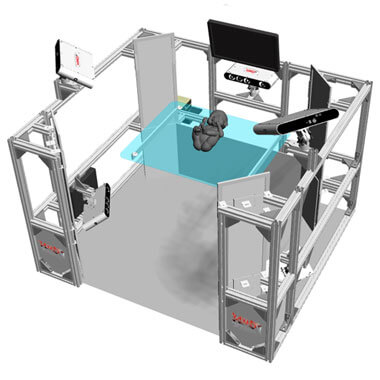
It contains 90k 3dMD system-captured RGB images and 13k 3dMDhand scans from 263 subjects (131 male, 132 female) with diverse skin tones and hand shapes.
For 20-plus years, 3dMD has enabled its worldwide customer community to successfully Train AI, Wear Tech, and Image Health. With a proven clinical background, 3dMD offers industrial-strength temporal-3D (4D-motion) dense-surface camera systems for human subject applications demanding ‘near ground truth’ 3D shape accuracy including healthcare, Computer Vision, Artificial Intelligence, Machine Learning, humanoid robotics, wearable-tech, human factors, ergonomics, anthropometrics, AR/VR/XR, etc. With a highly efficient subject workflow suitable for recruiting thousands of subjects, 3dMD supports high-volume head, face, full body, hand, and foot performance capture exercises for many customer innovations from clinical findings and ‘real world’ AI/ML training to groundbreaking design of wearable tech, performance apparel, and efficient ergonomics. 3dMD’s robust technology has sustained the findings in hundreds of peer-reviewed investigative research publications – many of which are found here on our website.
From Healthcare to Artificial Intelligence, here are a few 3dMD Applications…
Blog / News
Three-dimensional evaluation revealed facial soft tissue deformities in both groups, primarily in the nasolabial area.
Children with adenotonsillar hypertrophy and mouth breathing demonstrate narrowing of the nasopharyngeal, palatal, and glossopharyngeal regions.


