Be Part of the Alder Hey Children’s Hospital Headspace Project and Change Lives!

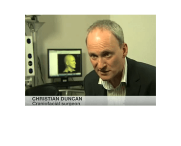
Liverpool, United Kingdom (24-September-13): Alder Hey Children’s Hospital and FACT are looking for volunteers to take part in a groundbreaking project which will impact on the future development of craniofacial surgery. The Headspace project, created by Alder Hey in conjunction with FACT and associated artists will develop a comprehensive database of human head shapes using…

Unilateral Condylar Hyperplasia: A 3-Dimensional Quantification of Asymmetry. TJ Verhoeven, JW Nolte, TJJ Maal, SJ Bergé, AG Becking.

Date: March 2013. Source: PLoS ONE.org. Purpose: Objective quantifications of facial asymmetry in patients with Unilateral Condylar Hyperplasia (UCH) have not yet been described in literature. The aim of this study was to objectively quantify soft-tissue asymmetry in patients with UCH and to compare the findings with a control group using a new method. Material…

Soft-Tissue Volumetric Changes Following Monobloc Distraction Procedure: Analysis Using Digital Three-Dimensional Photogrammetry System (3dMD). FC Chan, HK Kawamoto, C Federico, JP Bradley.

Date: March 2013. Source: Journal of Craniofacial Surgery, Volume 24, Issue 2, pp 416-420. Abstract: We have previously reported that monobloc advancement by distraction osteogenesis resulted in decreased morbidity and greater advancement with less relapse compared with acute monobloc advancement with bone grafting. In this study, we examine the three-dimensional (3D) volumetric soft-tissue changes in…

Three-Dimensional Analysis of Facial Asymmetry of Healthy Hispanic Caucasian Children. J Lee, B Ku, AC Da Silveira, MK Markey.

Date: October 16-17, 2012 Source: 3rd International Conference on 3D Body Scanning Technologies 2012, Lugano, Switzerland Abstract: The objective of this study was to conduct a quantitative analysis of facial asymmetry of healthy Hispanic Caucasian children residing in Central Texas, USA. 3D facial surface images of healthy Hispanic Caucasian participants (41 boys, 39 girls) currently…

Computation of an average cranial shape of three to twelve months old infants using 3D stereophotogrammetry. T Maal, L Verhamme, E van Lindert, W Borstlap, S Berge.

Date: June 28, 2012. Source: 26th International Congress of Computer Assisted Radiology 2012 (CARS) in Pisa, Italy. Purpose: The goal of this study was to create an average cranial shape using an automated process for a control population. This model includes the variation of growth and allows for an easy comparison between patient and average…

One year postoperative hard and soft tissue volumetric changes after a BSSO mandibular advancement. TJJ Maal, MJJ de Koning, JM Plooij, LM Verhamme, FA Rangel, SJ Bergé, WA Borstlap.

Date: May 2012 Source: International Journal of Oral and Maxillofacial Surgery; Volume 41(9), pp 1137-45. Abstract: In this study, cone beam computed tomography (CBCT) and three dimensional (3D) stereophotogrammetry are used to compare the 3D skeletal and soft tissue changes caused by a bilateral sagittal split osteotomy (BSSO) 1 year after a mandibular advancement. Eighteen…

Shape-Based Classification of 3D Facial Data to Support 22q11.2DS Craniofacial Research. K Wilamowska, J Wu, C Heike and L Shapiro

Date: March 2012 Source: Journal of Digital Imaging, Volume 25, Number 3 (2012), 400-408. Abstract: 3D imaging systems are used to construct high-resolution meshes of patient’s heads that can be analyzed by computer algorithms. Our work starts with such 3D head meshes and produces both global and local descriptors of 3D shape. Since these descriptors…

Variation of the Face in Rest Using 3D Stereophotogrammetry. TJJ Maal, LM Verhamme, B van Loon, JM Plooij, A Kho, FA Rangel, W Borstlap, SJ Bergé

Date: November 2011. Source: International Journal of Oral and Maxillofacial Surgery, Volume 40, Issue 11, Pages 1252-1257. Abstract: To evaluate treatment outcomes following oral and maxillofacial surgery, pre- and post-treatment three-dimensional (3D) photographs of the patient’s face can assessed, but this procedure is accurate only if the face is captured with the same facial expression…

Facial phenotypes in subgroups of prepubertal boys with autism spectrum disorders are correlated with clinical phenotypes. K Aldridge, ID George, KK Cole, JR Austin, TN Takahashi, Y Duan, and JH Miles.

Date: October 2011. Source: Molecular Autism Journal, 2:15. Background: The brain develops in concert and in coordination with the developing facial tissues, with each influencing the development of the other and sharing genetic signaling pathways. Autism spectrum disorders (ASDs) result from alterations in the embryological brain, suggesting that the development of the faces of children…