Detecting Genetic Association of Common Human Facial Morphological Variation Using High Density 3D Image Registration. S Peng, J Tan, S Hu, H Zhou, J Guo, L Jin, K Tang.

Date: December 2013. Source: PLoS Computational Biology, Public Library of Science. Abstract: Human facial morphology is a combination of many complex traits. Little is known about the genetic basis of common facial morphological variation. Existing association studies have largely used simple landmark-distances as surrogates for the complex morphological phenotypes of the face. However, this can…

BBC: Defining the most 'normal' shape of a human skull.

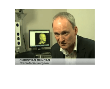
Date: 22 November 2013 Source: BBC News Article: Defining the most ‘normal’ shape of a human skull Reconstructive surgery for people born with a misshapen skull is one of medicine’s most complicated challenges. Part of the problem for surgeons is defining what a normal head actually looks like.  A team of Liverpool-based surgeons, and artists,…

Indiana University visualization experts help doctors diagnose fetal alcohol spectrum disorders with 3dMD

Date: October 10, 2013 Source: Indiana University, IT News & Events BLOOMINGTON, Ind. — Indiana University received a $125,000 grant renewal from the National Institutes of Health for ongoing research into detecting fetal alcohol spectrum disorders, a leading but preventable cause of birth defects. IU has been engaged in the Collaborative Initiative on Fetal Alcohol…

Be Part of the Alder Hey Children’s Hospital Headspace Project and Change Lives!

Liverpool, United Kingdom (24-September-13): Alder Hey Children’s Hospital and FACT are looking for volunteers to take part in a groundbreaking project which will impact on the future development of craniofacial surgery. The Headspace project, created by Alder Hey in conjunction with FACT and associated artists will develop a comprehensive database of human head shapes using…

Comparison of facial morphologies between adult Chinese and Houstonian Caucasian populations using three-dimensional imaging. J Wirthlin, CH Kau, JD English, F Pan, H Zhou.

Date: Sep 2013 Source: International Journal of Oral and Maxillofacial Surgery, 42(9):1100-7. Abstract: The objective of this study was to compare the facial morphologies of an adult Chinese population to a Houstonian white population. Three-dimensional (3D) images were acquired via a commercially available stereophotogrammetric camera system, 3dMDface™. Using the system, 100 subjects from a Houstonian…

Image Fusion in Craniofacial Virtual Reality Modeling Based on CT and 3dMD Photogrammetry. P Xin; H Yu; H Cheng; S Shen; SGF Shen.

Date: September 2013. Source: Journal of Craniofacial Surgery: Volume 24, Issue 5, pp 1573–1576. Purpose: The aim of this study was to demonstrate the feasibility of building a craniofacial virtual reality model by image fusion of 3-dimensional (3D) CT models and 3dMD stereophotogrammetric facial surface. Methods: A CT scan and stereophotography were performed. The 3D…

Presentation: Use of Digital Design. A Paradigm Shift in Surgical Planning for Craniofacial Surgery…Integrating Clinical Data with Static 3D Images. PR Shetye.

Date: September 2013. Source: International Society of Craniofacial Surgery 15th International Congress 2013, Jackson Hole, Wyoming, USA. Presentation: Use of Digital Design. A Paradigm Shift in Surgical Planning for Craniofacial Surgery… Integrating Clinical Data with Static 3D Images. Speaker: Pradip R. Shetye, DDS NYU Medical Center.

3-Dimensional Facial Simulation in Orthognathic Surgery: Is It Accurate? SA Schendel, R Jacobson, S Khalessi.

Date: Aug 2013. Source: Journal of Oral and Maxillofacial Surgery, 71(8):1406-14. Purpose: The purpose of this study was to measure the accuracy of 3D computer simulation of soft tissue changes after orthognathic surgery. Materials and Methods: Consecutive patients who underwent orthognathic surgery were studied by photogrammetric facial scanning and cone-beam computed tomography before and after…

BBC News: Cardiff University uses 3D scans to help facial surgery.

Date: 2 August 2013 Source: BBC News News: Cardiff University uses 3D scans to help facial surgery. Patients who need facial surgery and those recovering from strokes could benefit from world-leading research of analysing facial movements in 3D. Scientists at Cardiff University are using cutting-edge face scanners developed by American firm 3dMD. The technology can…

Cardiff Scientists and 3dMD Enter a New Dimension in 3D/4D Facial Imaging.

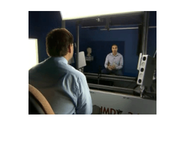
Anaheim, California (23-July-13): A team of Cardiff University clinicians and computer scientists can, for the first time, carry out facial movement research and transform the way patients needing facial surgery are assessed and monitored thanks to a new state-of-the-art 3D surface motion imaging system from 3dMD (3dMD exhibiting at #SIGGRAPH2013 Booth 226). Working alongside the…