Image Fusion in Preoperative Planning. SA Schendel, KS Duncan, CR Lane.

Date: November 2011. Source: 3D Imaging Technologies for Facial Plastic Surgery, Facial Plastic Surgery Clinics of North America, Vol 19, Issue 4 , Pages 577-590. Abstract: This article presents a comprehensive overview of generating a digital Patient-Specific Anatomic Reconstruction (PSAR) model of the craniofacial complex as the foundation for a more objective surgical planning platform.…

Facial dimensions of Malay children with repaired unilateral cleft lip and palate: a three dimensional analysis. M Zreaqat, R Hassan, AS Halim.

Date: June 2012. Source: International Journal of Oral and Maxillofacial Surgery; 41(6):783-8. Abstract: This comparative cross-sectional study assessed the facial surface dimensions of a group of Malay children with unilateral cleft lip and palate (UCLP) and compared them with a control group. 30 Malay children with UCLP aged 8–10 years and 30 unaffected age-matched children…

Statistical modelling of lip movement in the clinical context. H Popat, AI Zhurov, AM Toma, S Richmond, D Marshall, PL Rosin.

Date: May 2012. Source: Orthodontics & Craniofacial Research; Volume 15, Issue 2, pp 92–102. Objective: To establish three-dimensional (3D) reference data on average lip movement in normal healthy subjects using statistical shape analysis techniques. Setting and Sample Population: School of Dentistry and Cardiff School of Computer Science, Cardiff University, United Kingdom. One hundred and fifteen…

Shape-Based Classification of 3D Facial Data to Support 22q11.2DS Craniofacial Research. K Wilamowska, J Wu, C Heike and L Shapiro

Date: March 2012 Source: Journal of Digital Imaging, Volume 25, Number 3 (2012), 400-408. Abstract: 3D imaging systems are used to construct high-resolution meshes of patient’s heads that can be analyzed by computer algorithms. Our work starts with such 3D head meshes and produces both global and local descriptors of 3D shape. Since these descriptors…

BBC Health. 3D Face Scan to Help Future Surgery.

Date: January 2012. Source: BBC Health. 3D face scan to help future surgery Article: Visitors to London’s Science Museum are being invited to have their faces scanned in 3D. The ‘Me in 3D’ exhibit uses an array of cameras to build a virtual image visitors can then view and manipulate. Data from the participants will…

BBC News: London's Science Museum to scan visitors' faces in 3D

Date: 17 January 2012. Source: BBC News: http://www.bbc.co.uk/news/health-16549098 Article: Visitors to London’s Science Museum are being invited to have their faces scanned in 3D. The Me in 3D stand at the museum uses a series of cameras to build a virtual image visitors can then view and manipulate. Data from participants will be used by…

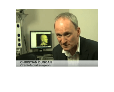
BBC News: 3D face scan to help future surgery.

Date: 17 January 2012. Source: BBC News: http://www.bbc.co.uk/news/science-environment-16588956 News Segment: 3D face scan to help future surgery. Visitors to London’s Science Museum are being invited to have their faces scanned in 3D. The ‘Me in 3D’ exhibit uses an array of cameras to build a virtual image visitors can then view and manipulate. Data from…

Announcement. See yourself in 3D at the London Science Museum.

Date: January 2012. Source: Press Announcement LONDON (3 January 2012) – How are our faces constructed? How does your face differ from other faces? What do you look like in another dimension? These questions will be explored in Me in 3D – a new event running from 11 January until 10 April, where visitors can…

Maxillary, mandibular, and chin advancement: treatment planning based on airway anatomy in obstructive sleep apnea. SA Schendel, N Powell, R Jacobson.

Date: March 2011 Source: Journal of Oral Maxillofacial Surgery, 69(3): 663-76. Abstract: Surgical correction of obstructive sleep apnea (OSA) syndrome involves understanding a number of parameters, of which the 3-dimensional airway anatomy is important. Visualization of the upper airway based on cone beam computed tomography scans and automated computer analysis is an aid in understanding…

3dMD is awarded Phase I of STTR Grant with UNC at Chapel Hill for a Dynamic 4D Facial Soft Tissue Analysis System.

Date: March 2011 Source: Press Announcement Headline: 3dMD is awarded Phase I of STTR Grant No. 1R41DE019742-01A1 with the University of North Carolina at Chapel Hill, Co-Principal Investigator Dr Carroll Ann Trotman, for a Dynamic 4D Facial Soft Tissue Analysis System. Atlanta (15-March-11): We are delighted to announce that 3dMD, working in conjunction with the Department of…