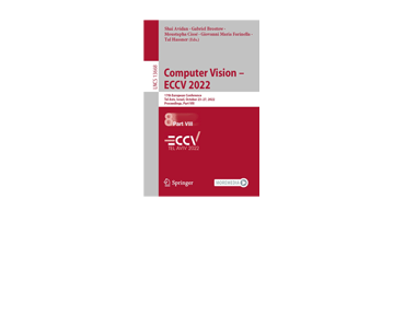
MimicME: A Large Scale Diverse 4D Database for Facial Expression Analysis. A Papaioannou et al.
Imperial College London captures real-world MimicMe, a 4,700-subject 4D facial expression database, with 3dMD, presented at ECCV 2022.
Training AI, Wearing Tech,
and Imaging Health.

Imperial College London captures real-world MimicMe, a 4,700-subject 4D facial expression database, with 3dMD, presented at ECCV 2022.
This excerpt will show up on the home page Blog/News feed

Imperial College London and Huawei build a 3D tongue reconstruction model with 3dMD’s real-world Science Museum, London dataset.

Great Ormond Street Hospital, Imperial College London, and Alder Hey use 3dMD data to train a 3D shape-based research model for analyzing craniofacial syndromes.

The Khronos Group is creating interoperability standards for 3D, AR/VR, vision, and machine learning.

Meta Reality Labs and Imperial College London build a neural hand deformation model with 3dMD’s real-world 3DH scan data, 2021.

Alder Hey and University of York use 3dMD’s real-world Headspace dataset to model craniofacial growth and sexual dimorphism in children, using geometric morphometrics.
University of York and Alder Hey use 3dMD’s real-world Headspace dataset to model craniofacial aging across adulthood, finding men and women age in distinct, non-linear patterns.
The proposed method was evaluated on the analysis of 3D facial shape and patient characteristics (N=5011). Experiments showed that this conceptually simple method achieved state-of-the-art fair prediction performance and interpretability, showing its great potential for clinical applications.

This excerpt will show up on the home page Blog/News feed