Craniofacial Image Analysis. E Mercan, I Atmosukarto, J Wu, S Liang, LG Shapiro.

Date: July 2015. Source: Health Monitoring and Personalized Feedback using Multimedia Data. Springer. Abstract: Craniofacial researchers have used anthropometric measurements taken directly on the human face for research and medical practice for decades. With the advancements in 3D imaging technologies, computational methods have been developed for the diagnoses of craniofacial syndromes and the analysis of…

The Use of 3D Imaging in Craniosynostosis; Surgical Planning; and Follow-Up. T Maal.

Date: June 24, 2015. Source: European Craniofacial Congress 2015, Gothenburg, Sweden. Presentation: The Use of 3D Imaging in Craniosynostosis; Surgical Planning; and Follow-Up. Presenter: Thomas J Maal, 3D Lab, Department of Oral- and Maxillofacial Surgery, Radboud University Medical Centre, Nijmegen, Netherlands.

Methods for Extraction and Analysis of Shape and Asymmetry from Craniofacial 3D Images. TA Darvann.

Date: June 24, 2015. Source: European Craniofacial Congress 2015, Gothenburg, Sweden. Summary: Being able to compare images is one of the core technologies of medical image analysis. In order to quantify change, e.g. due to growth or surgery, it is necessary to compare two or more images. And as part of population studies it is…

A Solution to Face-to-Face Contact in Tele-Presence Systems. X Zhou, P Boulanger.

Date: June 2015 Source: 2015 12th Conference on Computer and Robot Vision (CRV) Proceedings (p 230 – 236), Halifax, Canada. Abstract: In tele-presence systems, direct face-to-face contact, is essential for emotional communication as it carries non-verbal clues. In commercial products, face-to-face contact is approximated by using either a large semi-transparent mirror to project an image…

Thesis: Investigating the Accuracy and Reproducibility of 3dMDface System for Soft Tissue Analysis. YM Kachroo.

Date: June 2015. Source: Theses. ProQuest, Publication No. 1590412. University of California, Los Angeles. Advisers: Yeumin Hong; Won Moon. Objective: Three-dimensional (3D) soft tissue (ST) changes from orthodontic treatment can now be evaluated with the development of 3D photography. However, the accuracy and reliability of this method is still in question. This study aims to…

Three-dimensional human facial morphologies as robust aging markers. W Chen, W Qian, G Wu, W Chen, B Xian, X Chen, Y Cao, CD Green, F Zhao, K Tang, JD Han.

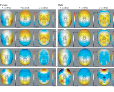
Date: May 2015. Source: Journal of Cell Research, 25, 574-587. Abstract: Aging is associated with many complex diseases. Reliable prediction of the aging process is important for assessing the risks of aging-associated diseases. However, despite intense research, so far there is no reliable aging marker. Here we addressed this problem by examining whether human 3D…

3D surface imaging of the human female torso in upright to supine positions. GP Reece, F Merchant, J Andon, H Khatam, K. Ravi-Chandar, J Weston, MC Fingeret, CR Lane, KS Duncan, MK Markey.

Date: April 2015. Source: Medical, Engineering and Physics, Volume 37, Issue 4, Pages 375–383. Abstract: Three-dimensional (3D) surface imaging of breasts is usually done with the patient in an upright position, which does not permit comparison of changes in breast morphology with changes in position of the torso. In theory, these limitations may be eliminated if…

Rating nasolabial appearance on three-dimensional images in cleft lip and palate: a comparison with standard photographs. A Stebel, D Desmedt, E Bronkhorst, MA Kuijpers, PS Fudalej.

Date: April 2015 (online). Source: The European Journal of Orthodontics. Background/Objective: Judgement of nasolabial aesthetics in cleft lip and palate (CLP) is a vital component of assessment of treatment outcome. It is usually performed based on two-dimensional (2D) facial photographs. An increasing use of three-dimensional (3D) imaging warrants an assessment if 3D images can substitute…

Monitoring of Therapy for Mucopolysaccharidosis Type I Using Dysmorphometric Facial Phenotypic Signatures. Stefanie Kung, M Walters, P Claes, P LeSouef, J Goldblatt, A Martin, S Balasubramaniam, G Baynam.

Date: March 2015. Source: JIMD Reports 2015, Springer. Abstract: There is a pattern of progressive facial dysmorphology in mucopolysaccharidosis type I (MPS I). Advances in 3D facial imaging have facilitated the development of tools, including dysmorphometrics, to objectively and precisely detect these facial phenotypes. Therefore, we investigated the application of dysmorphometrics as a noninvasive therapy-monitoring…