Creation of the Virtual Patient for the Study of Facial Morphology. CH Kau.

Date: November 2011. Source: 3D Imaging Technologies for Facial Plastic Surgery, Facial Plastic Surgery Clinics of North America, Volume 19, Number 4, Pages 615-622. Abstract: The author provides an overview of the new imaging technologies that allow the practitioner to accurately capture the patient’s soft tissue facial morphology and underlying bones and teeth, including details…

Image Fusion in Preoperative Planning. SA Schendel, KS Duncan, CR Lane.

Date: November 2011. Source: 3D Imaging Technologies for Facial Plastic Surgery, Facial Plastic Surgery Clinics of North America, Vol 19, Issue 4 , Pages 577-590. Abstract: This article presents a comprehensive overview of generating a digital Patient-Specific Anatomic Reconstruction (PSAR) model of the craniofacial complex as the foundation for a more objective surgical planning platform.…

Facial dimensions of Malay children with repaired unilateral cleft lip and palate: a three dimensional analysis. M Zreaqat, R Hassan, AS Halim.

Date: June 2012. Source: International Journal of Oral and Maxillofacial Surgery; 41(6):783-8. Abstract: This comparative cross-sectional study assessed the facial surface dimensions of a group of Malay children with unilateral cleft lip and palate (UCLP) and compared them with a control group. 30 Malay children with UCLP aged 8–10 years and 30 unaffected age-matched children…

One year postoperative hard and soft tissue volumetric changes after a BSSO mandibular advancement. TJJ Maal, MJJ de Koning, JM Plooij, LM Verhamme, FA Rangel, SJ Bergé, WA Borstlap.

Date: May 2012 Source: International Journal of Oral and Maxillofacial Surgery; Volume 41(9), pp 1137-45. Abstract: In this study, cone beam computed tomography (CBCT) and three dimensional (3D) stereophotogrammetry are used to compare the 3D skeletal and soft tissue changes caused by a bilateral sagittal split osteotomy (BSSO) 1 year after a mandibular advancement. Eighteen…

Evaluation of direct and indirect additive manufacture of maxillofacial prostheses. D Eggbeer, R Bibb, P Evans, L Ji

Date: April 2012. Source: Journal Of Engineering in Medicine, vol. 226 no. 9 718-728. Abstract: The efficacy of computer-aided technologies in the design and manufacture of maxillofacial prostheses has not been fully proven. This paper presents research into the evaluation of direct and indirect additive manufacture of a maxillofacial prosthesis against conventional laboratory-based techniques. An…

BBC News: London's Science Museum to scan visitors' faces in 3D

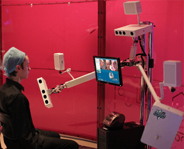
Date: 17 January 2012. Source: BBC News: http://www.bbc.co.uk/news/health-16549098 Article: Visitors to London’s Science Museum are being invited to have their faces scanned in 3D. The Me in 3D stand at the museum uses a series of cameras to build a virtual image visitors can then view and manipulate. Data from participants will be used by…

Announcement. See yourself in 3D at the London Science Museum.

Date: January 2012. Source: Press Announcement LONDON (3 January 2012) – How are our faces constructed? How does your face differ from other faces? What do you look like in another dimension? These questions will be explored in Me in 3D – a new event running from 11 January until 10 April, where visitors can…

Variation of the Face in Rest Using 3D Stereophotogrammetry. TJJ Maal, LM Verhamme, B van Loon, JM Plooij, A Kho, FA Rangel, W Borstlap, SJ Bergé

Date: November 2011. Source: International Journal of Oral and Maxillofacial Surgery, Volume 40, Issue 11, Pages 1252-1257. Abstract: To evaluate treatment outcomes following oral and maxillofacial surgery, pre- and post-treatment three-dimensional (3D) photographs of the patient’s face can assessed, but this procedure is accurate only if the face is captured with the same facial expression…

Image Fusion in Preoperative Planning. SA Schendel, KS Duncan, C Lane.

Date: November 2011. Source: 3D: Fusion, Sculpting, Imaging Systems. Facial Plastic Surgery Clinics of North America. Volume 19, Issue 4, Pages 577-590. Abstract: This article presents a comprehensive overview of generating a digital Patient-Specific Anatomic Reconstruction (PSAR) model of the craniofacial complex as the foundation for a more objective surgical planning platform. The technique explores…

Maxillary, mandibular, and chin advancement: treatment planning based on airway anatomy in obstructive sleep apnea. SA Schendel, N Powell, R Jacobson.

Date: March 2011 Source: Journal of Oral Maxillofacial Surgery, 69(3): 663-76. Abstract: Surgical correction of obstructive sleep apnea (OSA) syndrome involves understanding a number of parameters, of which the 3-dimensional airway anatomy is important. Visualization of the upper airway based on cone beam computed tomography scans and automated computer analysis is an aid in understanding…