Digital imaging analysis to assess scar phenotype. BJ Smith, N Nidey, SF Miller, LMM Uribe, CL Baum, GS Hamilton, GL Wehby, M Dunnwald.

Date: March-April 2014. Source: Wound Repair and Regeneration, Volume 22, Issue 2, pages 228–238. Abstract: In order to understand the link between the genetic background of patients and wound clinical outcomes, it is critical to have a reliable method to assess the phenotypic characteristics of healed wounds. In this study, we present a novel imaging…

Three-Dimensional Stereophotogrammetry: A Novel Method in Volumetric Measurement of Infantile Hemangioma. DJJ Hermans, TJJ Maal, SJ Bergé, CJM Vleuten.

Date: January 2014. Source: Pediatric Dermatology. Volume 31, Issue 1, pp 118-122. Abstract: Accurate and objective measurement of volume changes in infantile hemangiomas (IHs) is essential in routine clinical practice and clinical studies, particularly in the changing therapeutic landscape after the discovery of propranolol. Several bedside techniques for volume measurement have been described in the…

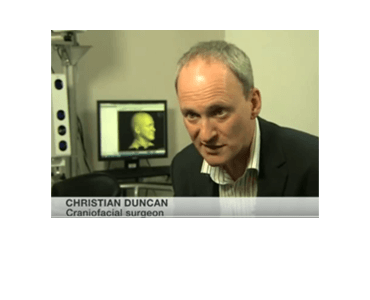
BBC: Defining the most 'normal' shape of a human skull.

Date: 22 November 2013 Source: BBC News Article: Defining the most ‘normal’ shape of a human skull Reconstructive surgery for people born with a misshapen skull is one of medicine’s most complicated challenges. Part of the problem for surgeons is defining what a normal head actually looks like.  A team of Liverpool-based surgeons, and artists,…

Be Part of the Alder Hey Children’s Hospital Headspace Project and Change Lives!

Liverpool, United Kingdom (24-September-13): Alder Hey Children’s Hospital and FACT are looking for volunteers to take part in a groundbreaking project which will impact on the future development of craniofacial surgery. The Headspace project, created by Alder Hey in conjunction with FACT and associated artists will develop a comprehensive database of human head shapes using…

Image Fusion in Craniofacial Virtual Reality Modeling Based on CT and 3dMD Photogrammetry. P Xin; H Yu; H Cheng; S Shen; SGF Shen.

Date: September 2013. Source: Journal of Craniofacial Surgery: Volume 24, Issue 5, pp 1573–1576. Purpose: The aim of this study was to demonstrate the feasibility of building a craniofacial virtual reality model by image fusion of 3-dimensional (3D) CT models and 3dMD stereophotogrammetric facial surface. Methods: A CT scan and stereophotography were performed. The 3D…

Presentation: Use of Digital Design. A Paradigm Shift in Surgical Planning for Craniofacial Surgery…Integrating Clinical Data with Static 3D Images. PR Shetye.

Date: September 2013. Source: International Society of Craniofacial Surgery 15th International Congress 2013, Jackson Hole, Wyoming, USA. Presentation: Use of Digital Design. A Paradigm Shift in Surgical Planning for Craniofacial Surgery… Integrating Clinical Data with Static 3D Images. Speaker: Pradip R. Shetye, DDS NYU Medical Center.

3-Dimensional Facial Simulation in Orthognathic Surgery: Is It Accurate? SA Schendel, R Jacobson, S Khalessi.

Date: Aug 2013. Source: Journal of Oral and Maxillofacial Surgery, 71(8):1406-14. Purpose: The purpose of this study was to measure the accuracy of 3D computer simulation of soft tissue changes after orthognathic surgery. Materials and Methods: Consecutive patients who underwent orthognathic surgery were studied by photogrammetric facial scanning and cone-beam computed tomography before and after…

Fat graft transfer in children's facial malformations: A prospective three-dimensional evaluation. M Guibert, G Franchi, E Ansari, B Billotet, PA Diner, S Cassier, MP Vazquez, A Picard, N Kadlub.

Date: June 2013. Source: Journal of Plastic, Reconstructive & Aesthetic Surgery, 66(6):799-804. Introduction: Autologous fat grafting for volume augmentation in soft tissues has grown in popularity in the paediatric plastic surgery community, despite a lack of quantifiable evidence of graft survival and predictable outcomes for child patients. The objective of this study is to quantify,…

Three-dimensional evaluation of unilateral cleft rhinoplasty results. TK Dixon, BP Caughlin, N Munaretto, DM Toriumi.

Date: April 2013 Source: Facial Plastic Surgery.; 29(2): 106-15. doi: 10.1055/s-0033-1341588. Abstract: Three-dimensional (3D) imaging is a relatively new method of objectively evaluating surgical results, allowing the surgeon to accurately measure postsurgical changes with little inconvenience to the patient. Its accuracy and reliability has been consistently demonstrated in the literature. This article describes updated methods…