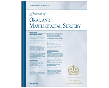
3-Dimensional Facial Simulation in Orthognathic Surgery: Is It Accurate? SA Schendel, R Jacobson, S Khalessi.

Date: Aug 2013. Source: Journal of Oral and Maxillofacial Surgery, 71(8):1406-14. Purpose: The purpose of this study was to measure the accuracy of 3D computer simulation of soft tissue changes after orthognathic surgery. Materials and Methods: Consecutive patients who underwent orthognathic surgery were studied by photogrammetric facial scanning and cone-beam computed tomography before and after…

BBC News: Cardiff University uses 3D scans to help facial surgery.

Date: 2 August 2013 Source: BBC News News: Cardiff University uses 3D scans to help facial surgery. Patients who need facial surgery and those recovering from strokes could benefit from world-leading research of analysing facial movements in 3D. Scientists at Cardiff University are using cutting-edge face scanners developed by American firm 3dMD. The technology can…

Cardiff Scientists and 3dMD Enter a New Dimension in 3D/4D Facial Imaging.

Anaheim, California (23-July-13): A team of Cardiff University clinicians and computer scientists can, for the first time, carry out facial movement research and transform the way patients needing facial surgery are assessed and monitored thanks to a new state-of-the-art 3D surface motion imaging system from 3dMD (3dMD exhibiting at #SIGGRAPH2013 Booth 226). Working alongside the…

Improved Detection of Landmarks on 3D Human Face Data. S Liang; S Weinberg; L Shapiro.

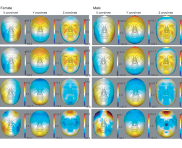
Date: July 2013. Source: IEEE Engineering in Medicine and Biology Society Annual Conference. Abstract: Craniofacial researchers make heavy use of established facial landmarks in their morphometric analyses. For studies on very large facial image datasets, the standard approach of manual landmarking is very labor intensive. With the goal of producing 20 established landmarks, we have…

The Use of Pseudo-landmarks for Craniofacial Analysis: A Comparative Study with L1 Regularized Logistic Regression. E Mercan; L Shapiro; S Weinberg; SI Lee.

Date: July 2013. Source: IEEE Engineering in Medicine and Biology Society Annual Conference. Abstract: Morphometrics, the quantitative analysis of shape, is used by craniofacial researchers to study abnormalities in human face shapes. Most of the work in craniofacial morphometrics uses landmark points that are manually marked on 3D face data and processed via a generalized…

Fat graft transfer in children's facial malformations: A prospective three-dimensional evaluation. M Guibert, G Franchi, E Ansari, B Billotet, PA Diner, S Cassier, MP Vazquez, A Picard, N Kadlub.

Date: June 2013. Source: Journal of Plastic, Reconstructive & Aesthetic Surgery, 66(6):799-804. Introduction: Autologous fat grafting for volume augmentation in soft tissues has grown in popularity in the paediatric plastic surgery community, despite a lack of quantifiable evidence of graft survival and predictable outcomes for child patients. The objective of this study is to quantify,…

Flat Fold and Cup-Shaped N95 Filtering Facepiece Respirator Face Seal Area and Pressure Determinations: A Stereophotogrammetry Study. G Niezgoda, JH Kim, RJ Roberge, SM Benson.

Date: May 2013. Source: Journal of Occupational and Environmental Hygiene, Volume 10, Issue 8, pp 419-424. Abstract: Twenty subjects underwent quantitative respirator fit testing with two styles (flat fold, cup-shaped) of N95 filtering facepiece respirators (N95 FFRs). Passing a fit test was followed by stereophotogrammetry to determine the face seal area and computation of seal…

Three-dimensional reproducibility of natural head position. DW Weber, DW Fallis, MD Packerc.

Date: May 2013 Source: American Journal of Orthodontics and Dentofacial Orthopedics, Volume 143, Issue 5, pp 738-744. Introduction: Although natural head position has proven to be reliable in the sagittal plane, with an increasing interest in 3-dimensional craniofacial analysis, a determination of its reproducibility in the coronal and axial planes is essential. This study was…