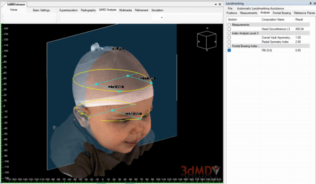
3D Photography to Quantify the Severity of Metopic Craniosynostosis. MK Bruce, WH Tao, J Beiriger, C Christensen, MJ Pfaff, R Whitaker, JA Goldstein.

Results of this study show that 3dMD photography is a valid alternative to CT for evaluation of head shape in MCS. Its use will provide an objective, quantifiable means of assessing outcomes in a rigorous manner while decreasing radiation exposure in this patient population.

School-age outcomes in patients with unilateral coronal synostosis: comparison of fronto-orbital advancement and endoscopic strip craniectomy. K Varagur, GB Skolnick, SD Naidoo, MD Smyth, KB Patel.

This study examines facial asymmetry, desire for revision, and patient-reported outcomes at school age in children who received either endoscopic strip craniectomy with helmet therapy or fronto-orbital advancement.

A Three-Dimensional-Based Morphometric Analysis of a Standardized Overcorrection Technique for Fronto-Orbital Advancement in Metopic Craniosynostosis. MJ Pfaff, MK Bruce, S Erpenbeck, et al.

he purpose of this study is to describe in detail a standardized technique to fronto-orbital advancement utilizing the concept of “overcorrection” and objectively evaluate intermediate results.

A Novel Study on Alar Mobility of HAN Female by 3dMD Dynamic Surface Imaging System. Y Zhong, Y Zhu, T Jiang et al.

Asian alar anatomy has great distinction from Caucasian, processing conspicuous alar movement and damaging alar aesthetic dynamically. This novel study defined alar mobility by three-dimensional anthropometric analysis, providing objective references for alar dynamic aesthetic and arousing plastic surgeons’ attention on keeping balance of static and dynamic aesthetic in rhinoplasty.

Three-Dimensional Nasolabial Changes After Nasoalveolar Molding and Primary Lip/Nose Surgery in Infants With Bilateral Cleft Lip and Palate. L Mancini, S Avinoam, BH Grayson, RL Flores, DA Staffenberg, PR Shetye.

This is a retrospective serial longitudinal study of consecutively enrolled infants from September 2012 to July 2016 with BCLP who underwent NAM before primary lip and nose reconstructive surgery.

Automatic Assessment of 3-Dimensional Facial Soft Tissue Symmetry Before and After Orthognathic Surgery Using a Machine Learning Model. LJ Lo, CT Yang, CT Ho, CH Liao, HH Lin.

This study applied the transfer learning model with a convolutional neural network based on 3-dimensional (3D) contour line features to evaluate the facial symmetry before and after OGS. A total of 158 patients were recruited in a retrospective cohort study for the assessment and comparison of facial symmetry before and after OGS from January 2018 to March 2020. Three-dimensional facial photographs were captured by the 3dMD face system in a natural head position, with eyes looking forward, relaxed facial muscles, and habitual dental occlusion before and at least 6 months after surgery.