Molecular genetic analysis in 14 czech kabuki syndrome patients is confirming the utility of phenotypic scoring. J Paděrová; A Holubová; M Simandlová; A Puchmajerová; M Vlčková; M Malíková; R Pourová; S Vejvalková; M Havlovicová; M Šenkeříková; N Ptáková; J Drábová; J Geryk; A Maver; A Křepelová; M Macek.

Date: February 2016. Source: Clinical Genetics. The International Journal of Genetics, Molecular, and Personalized Medicine. Abstract: Kabuki syndrome (KS) is a dominantly inherited disorder mainly due to de novo pathogenic variation in KMT2D or KDM6A genes. Initially, a representative cohort of 14 Czech cases with clinical features suggestive of KS was analyzed by experienced clinical…

Facial morphology in children and adolescents with juvenile idiopathic arthritis and moderate to severe temporomandibular joint involvement. YJ Hsieh, TA Darvann, NV Hermann, P Larsen, YF Liao, J Bjoern-Joergensen, S Kreiborg.

Date: February 2016 Source: American Journal of Orthodontics & Dentofacial Orthopedics. Volume 149, Issue 2, Pages 182–191. Highlights: • We evaluated lateral facial morphology of juvenile idiopathic arthritis patients with moderate and severe TMJ involvement. • Differences in their lateral facial morphologies between unilateral and bilateral TMJ involvement were also assessed. Introduction: The aims of…

Analysis of facial muscle activation in children with autism using 3D imaging. MD Samad, JL Bobzien, JW Harrington, KM Iftekharuddin.

Date: November 2015. Source: 2015 IEEE International Conference on Bioinformatics and Biomedicine (BIBM), Pages 337-342. Washington, DC. Abstract: Autism Spectrum Disorder (ASD) impairs an individual’s non-verbal skills including natural and contextual facial expressions. Such impairments may manifest as odd facial expressions (facial oddity) based on subjective evaluations of facial images. A few studies conducted on…

Analysis of Characteristic Facial Features in Patients with Hypodontia using Three-Dimensional Imaging. S Ani, J Collins, S Cunningham, C Ruff.

Date: September 2015. Source: 8th International Orthodontic Congress 2015, London. Study Aims: To analyse the facial features associated with hypodontia using 3D surface imaging and establish the effect of its severity on facial morphology. Method: A study group of 70 hypodontia patients and a control group of 40 Class I patients were recruited to allow…

Craniofacial Image Analysis. E Mercan, I Atmosukarto, J Wu, S Liang, LG Shapiro.

Date: July 2015. Source: Health Monitoring and Personalized Feedback using Multimedia Data. Springer. Abstract: Craniofacial researchers have used anthropometric measurements taken directly on the human face for research and medical practice for decades. With the advancements in 3D imaging technologies, computational methods have been developed for the diagnoses of craniofacial syndromes and the analysis of…

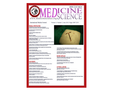
Comparison of the Facial Morphologies of Identical Twins Using Three-Dimensional Photography: A Case Report. Sedat Altindis, Erdem Hatunoglu, Emine Toptan.

Date: July 2015 Source: Medicine Science | International Medical Journal. Abstract: The facial morphologies of identical twins were compared using the 3dMD three-dimensional (3D) photogrammetry system. 3D images of the faces of 27-year-old identical twins were acquired and then superimposed. The differences were shown in a color histogram generated using 3dMD Vultus software. The faces…

Unilateral Cleft Lip Repair Using the Anatomical Subunit Approximation: Modifications and Analysis of Early Results in 100 Consecutive Cases. R Tse, S Lien.

Date: July 2015. Source: Plastic and Reconstructive Surgery, 136(1):119-30. Abstract: The “Anatomic Subunit Approximation” for unilateral cleft lip repair has gained acceptance, however, outcomes have not been reported since the original description. The purpose of this study is to report the experience using this technique. Methods: One hundred two consecutive patients underwent primary cleft lip…

Methods for Extraction and Analysis of Shape and Asymmetry from Craniofacial 3D Images. TA Darvann.

Date: June 24, 2015. Source: European Craniofacial Congress 2015, Gothenburg, Sweden. Summary: Being able to compare images is one of the core technologies of medical image analysis. In order to quantify change, e.g. due to growth or surgery, it is necessary to compare two or more images. And as part of population studies it is…

3D digital surface imaging for quantification of facial development and asymmetry in juvenile idiopathic arthritis. NV Hermann, TA Darvann, P Larsen, S Kreiborg.

Date: June 2015. Source: Seminars in Orthodontics, Volume 21, Issue 2, Pages 121–124. Abstract: 3D digital surface imaging (digital stereophotogrammetry or “3D photography”) is becoming an increasingly popular tool for quantification of the face, in clinical contexts as well as for research. The modality is easy to apply and is free from motion artifacts and…